Deep Back and Suboccipital Region
The muscles of the back are divided into extrinsic back muscles and intrinsic back muscles. The first two layers of muscles of the superficial back are extrinsic muscles primarily related to movements of the upper limb. The third layer of extrinsic back muscles are the serratus posterior muscles, which are thought to participate in respiration. These were dissected during the first lab.
The intrinsic (deep) back muscles are involved in axial movements, that is, movements related to the long axis of the vertebral column. In today’s dissection, we will examine the large deep back muscles (erector spinae), which extend (straighten) the flexed spine or, contracting unilaterally, bend the spine toward the same (ipsilateral) side. We will then examine the deeper small intrinsic back muscles. Additionally in this dissection, we will examine the suboccipital region located superiorly in the posterior part of the neck. It contains muscles that help produce movements of the head.
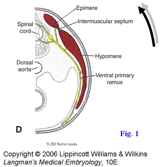
With the exception of the trapezius, the muscles that were studied in the dissection of the superficial back—latissimus dorsi, rhomboids, levator scapulae, and serratus posterior—all are innervated by the anterior (ventral) rami of spinal nerves (G11 1.18, 4.46B-C; G12 1.18, 4.49A; N 162A-B, 163, 174, 252). That is because they develop from the mass of embryonic mesoderm termed the hypomere (Fig. 1), which forms muscles of the extremities and body wall. The superficial back muscles originate elsewhere in the body of the embryo and migrate into the back, carrying their innervation from anterior rami with them.
The deep, or intrinsic, back muscles develop from a mass of embryonic mesoderm termed the epimere (Fig. 1) and are innervated by the posterior (dorsal) rami of spinal nerves. It will be necessary to cut and reflect the attachments of some deep back muscles in order to expose the suboccipital triangle.
The splenius capitis and splenius cervicis form the superficial layer of intrinsic back muscles in the neck and upper trunk. These two muscles have a continuous attachment to the lower part of the nuchal ligament (ligamentum nuchae) and the spinous processes of vertebrae Cv7 through Tv6 (G11 4.30, Table 4.4 [pp.314-315]; G12 4.33, Table 4.4-2-3; N168, 169). The more superior muscle, the splenius capitis, angles superolaterally to attach to the skull at the mastoid process of the temporal bone and along the lateral third of the superior nuchal line. The lower muscle, the splenius cervicis, attaches to the transverse processes of the upper three or four cervical vertebrae. Both muscles are segmentally innervated by the posterior rami of spinal nerves.
On a skeleton review the external occipital protuberance and the superior and inferior nuchal lines on the occipital bone of the skull and the mastoid process of the temporal bone (G11 7.2B, 7.3A, 7.4; G12 7.3B, 7.4A, 7.5; N6A-B, 10). Study the features of the first (atlas) and second (axis) cervical vertebrae (G11 4.10, 4.11; G12 4.9, 4.12A-B-C-D-E; N19A-B). The atlas and axis are atypical cervical vertebrae in which the body of the atlas essentially has been incorporated into the axis as a vertical process of bone, the dens. The atlas consists of two lateral masses joined by anterior and posterior arches, each with a midline tubercle on its external aspect. Each lateral mass has a transverse process passing laterally from it and bears superior and inferior articular surfaces/facets for articulation with an occipital condyle and the axis, respectively. Each transverse process contains a transverse foramen (foramen transversarium) for the vertebral artery of its side. There is also a groove for the vertebral artery on the superior surface of the posterior arch of the atlas on each side.
The axis resembles other cervical vertebrae except for the tooth-like process of bone, the dens or odontoid process, projecting superiorly from its body (N19A-B). The dens has an anterior articular facet for articulation with the anterior arch of the atlas and a posterior articular facet for articulation with the transverse ligament of the atlas (N23A-B). The transverse ligament of the atlas passes from one lateral mass of the atlas to the other and holds the dens against the anterior arch of the atlas. If the ligament is absent (e.g., in Down’s syndrome) or ruptures (e.g., in rheumatoid arthritis), atlantoaxial subluxation may occur with fatal cervical spinal cord injury.
Joints involving the atlas and axis provide much of the range of movement of the head. The atlas articulates superiorly with the occipital condyles of the skull at two atlanto-occipital joints to allow flexion and extension (as in nodding the head “yes”). The atlas and axis articulate with each other at three joints—one median and two lateral atlantoaxial joints (G11 Fig. 4.11C-D; G12 4.12C-D; N23A-B, 150). At the median atlantoaxial joint, the dens articulates anteriorly with the anterior arch of the atlas and posteriorly with the transverse ligament of the atlas, serving as a pivot around which the atlas (carrying the head) rotates, as in shaking the head “no.” The lateral atlantoaxial joints are formed between large inferior facets on the lateral masses of the atlas and superior articular processes on the axis. These lateral joints contribute to rotation.
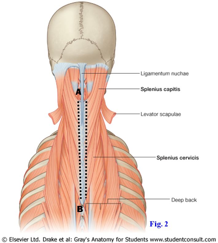
1. In the neck and upper trunk clean the splenius capitis and splenius cervicis. Cut the attachment of the splenius capitis and splenius cervicis from the nuchal ligament and spinous processes on each side and reflect the muscles laterally (Fig. 2, A-B).
Turn your attention to the next layer of deep back muscles on each side, the erector spinae (sacrospinalis) muscles (Fig. 3; G11 4.30, 4.31, Table 4.4 [pp. 314-315]; G12 4.33, 4.34, Table 4.4-2-3; N168, 169, 173). These muscles contract while shortening (concentric contraction) to return the trunk to an erect posture from a bent over, or flexed, position. They contract while lengthening (eccentric contraction) to control the rate and amount of flexion caused by another force (e.g., gravity). The erector spinae occupies the vertebrocostal groove deep to the posterior layer of thoracolumbar fascia (G11 4.31; G12 4.34; N173) and consists of three longitudinal columns of muscle, from lateral to medial the iliocostalis, longissimus, and spinalis. Each column is formed by muscle fascicles that span 6-10 segments between bony attachments (i.e., 6-10 ribs or vertebrae) and is subdivided into three regional parts.
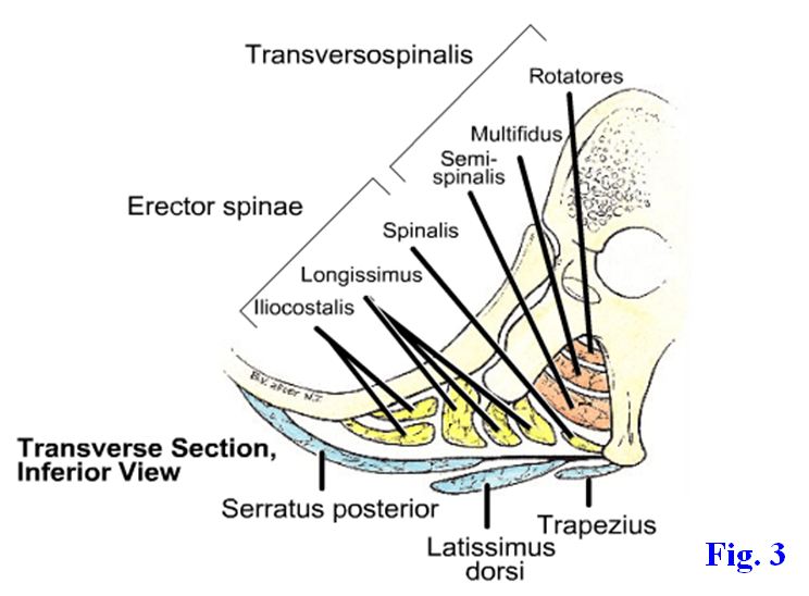
The lateral column of the erector spinae, the iliocostalis, basically ascends along the angles of the ribs (costal angles) and is divided into lumborum, thoracis, and cervicis parts (Fig. 3; G11 4.30; Table 4.4 pp. 314-315; G12 4.33, Table 4.4-2-3; N169) based on where the muscle is located. For example, the iliocostalis lumborum ascends from the sacrum and iliac crest (lumbar region) to the angles of the lower six or seven ribs; the iliocostalis thoracis ascends from the angles of the lower six ribs to the angles of the upper six ribs (thoracic region); the iliocostalis cervicis spans from the angles of ribs 3-6 to the transverse processes of Cv4 to Cv7 (cervical region).
The intermediate (middle) column, the longissimus, basically runs along the tips of the transverse processes of vertebrae and has thoracis, cervicis, and capitis parts (G11 4.30; Table 4.4 [pp. 314-315]; G12 4.33, Table 4.4-2-3; N169). Its capitis part reaches the skull to attach to the mastoid process. The boundary between the iliocostalis and longissimus columns of muscle basically is indicated by the emergence of the posterior rami of spinal nerves between them, although the rami can emerge through the lateral part of the longissimus (G11 4.30; G12 4.33).
The spinalis muscle is the most medial column of the erector spinae and runs along the sides of the spinous processes (G11 4.30; G12 4.33; N169). It has thoracis, cervicis, and capitis parts. The cervicis part is often absent. The capitis part is fused with the larger semispinalis capitis muscle and cannot be separated from it.
2. Clean the three columns of the erector spinae muscle. The iliocostalis is lateral, the longissimus is intermediate, and the spinalis is medial in position. Attempt to identify the regional subdivisions of each column. There are areas of overlap between the subdivisions (e.g., iliocostalis lumborum and iliocostalis thoracis), but identify areas in each column of erector spinae that clearly belong to only one subdivision.
The deep layer of intrinsic back muscles is the transversospinalis muscle, so called because the general orientation of the muscle fascicles is from the transverse processes of lower vertebrae to the spinous processes of more superior vertebrae. From superficial to deep, the transversospinalis consists of the semispinalis, multifidus, and rotatores (Fig. 3; G11 4.32-4.34; Table 4.4 [pp. 314-315]; G12 4.35, 4.36, 4.37, Table 4.4-2-3; N170). The semispinalis extends about half the length of the vertebral column, as the name implies, and its fascicles span 4-6 segments (e.g., from the transverse process of vertebra Tv10 to the spinous process of Tv5). The semispinalis has thoracis, cervicis, and capitis parts. The semispinalis cervicis muscles of the two sides converge to attach on the spinous process of the axis (Cv2) (G11 4.35, 4.36; G12 4.38, 4.39; N170). The semispinalis capitis is the substantial muscle responsible for the longitudinal bulge on the back of the neck near the midline. Its fibers attach on the occipital bone between the superior and inferior nuchal lines.
The multifidus lies deep to the semispinalis, and its fascicles span 2-4 segments. The multifidus is present from the sacrum to the spinous process of the axis but is most developed in the lumbar region (G11 4.33; G12 4.36; N170). The rotatores pass from a transverse process to the vertebra above (rotatores brevis) or span that vertebra to attach to the next one (rotatores longus) (i.e., the rotatores span 1-2 segments)(G11 4.34; G12 4.37). The rotatores are best developed in the thoracic region.
Small, minor muscles constitute the deepest group of back muscles and include the interspinales running between the spinous processes of vertebrae in the cervical and lumbar regions, intertransversarii connecting adjacent transverse processes, also in the cervical and lumbar regions, and the levatores costarum. The levatores costarum run from the tip of a transverse process down to the rib below, or sometimes to the second rib below.
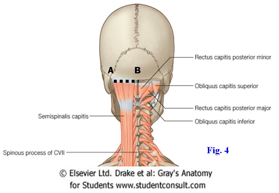
3. Clean the right and left semispinalis capitis muscles, which are separated in the midline by the fibroelastic nuchal ligament (G11 4.32, 4.35, Table 4.4 [p. 314]; G12 4.35, 4.38, Table 4.4-2-3; N22A-B [Right lateral view], 170). These thick, vertically oriented muscles are part of the transversospinalis(transversospinal)muscle group. Look for and preserve a large nerve piercing the semispinalis capitis and ascending into the scalp, the greater occipital nerve (G11 4.35; G12 4.38; N171, 172). This nerve is typically located about 2.5 cm from midline. The greater occipital nerve is the posterior ramus of spinal nerve C2 and pierces the semispinalis, and sometimes the trapezius, as it passes upward to provide cutaneous innervation to the posterior part of the scalp. Look for the occipital artery, a branch of the external carotid artery that ascends into the scalp just lateral to the greater occipital nerve.
4. Clean the semispinalis thoracis muscle. In the lumbar region attempt to demonstrate the multifidus. Remember that its fascicles span 2-4 segments in a superomedial direction from transverse to spinous processes. Remove a short segment of multifidus to expose examples of the rotatores.
THE SUBOCCIPITAL TRIANGLE
5. While protecting the greater occipital nerve, cut the attachment of the semispinalis capitis to the occipital bone (Fig. 4, A-B) and reflect the muscle laterally from the nuchal ligament and downward to about the level of Cv6 or Cv7 (G11 4.35, 4.36; G12 4.38, 4.39). You may have to follow the nerve through the upper part of the semispinalis capitis, cutting the muscle along the course of the nerve, in order to do this. Note the attachment of semispinalis cervicis muscles to the spinous process of the axis (Cv2).
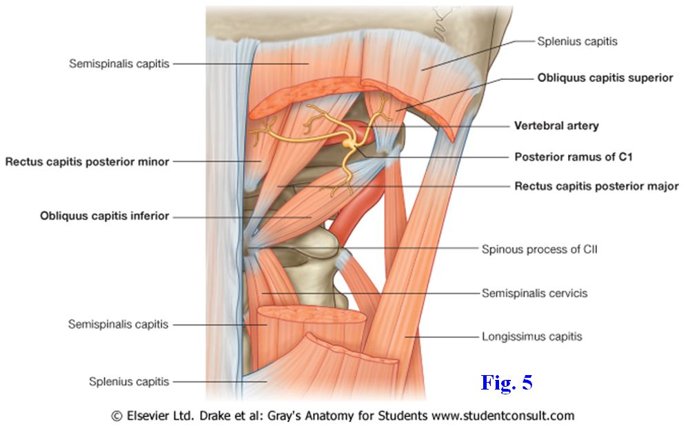
Reflection of the semispinalis capitis exposes the suboccipital triangle (Fig. 5; G11 4.36; G12 4.39; N170, 172) formed on each side by three short muscles that attach to the atlas and/or axis—rectus capitis posterior major, obliquus capitis inferior (inferior oblique of the head), and obliquus capitis superior (superior oblique of the head). The short rectus capitis posterior minor is located medial to the suboccipital triangle. These suboccipital muscles are all innervated by the posterior ramus of C1, the suboccipital nerve.
The rectus capitis posterior major passes from the spinous process of the axis (Cv2) to the lateral part of the inferior nuchal line, forming the medial boundary of the suboccipital triangle (G11 4.36, Table 4.5 [p. 318]; G12 4.39, Table 4.5-2-3; N170, 172). A smaller muscle, the rectus capitis posterior minor, passes from the posterior tubercle of the atlas (Cv1) to the medial part of the inferior nuchal line and lies medial to the triangle.
The obliquus capitis inferior muscle takes origin from the spinous process of the axis and inserts into the transverse process of the atlas (G11 4.36; G12 4.39; N170, 172). It forms the inferior boundary of the suboccipital triangle and is the only muscle with “capitis” in its name that doesn’t attach to the skull. The greater occipital nerve winds around the inferior border of the obliquus capitis inferior and ascends to pierce the semispinalis capitis en route to the posterior part of the scalp. The obliquus capitis superior muscle passes superiorly from the transverse process of the atlas to the occipital bone between the superior and inferior nuchal lines. The obliquus capitis superior forms the lateral boundary of the triangle.
A useful landmark in identifying the muscles of this region is the spinous process of the axis (Cv2). Attached to this spinous process are, from superior to inferior, the rectus capitis posterior major, the obliquus capitis inferior, and the semispinalis cervicis muscles on each side. Thus, the combination of the six muscles from both sides form a star-like arrangement that is easily located (Fig. 5; G11 4.36; G12 4.39; N172).6. Clean and identify the muscles of the suboccipital triangle (Fig. 5). Within the suboccipital triangle palpate the hard posterior arch of the atlas between two membranes, the posterior atlanto-occipital and posterior atlantoaxial membranes(G11 4.38B, Table 4.5 [p. 318], Posterior View; G12 4.13B, Table 4.5-2-3). On the superior surface of the posterior arch of the atlas clean the third, or horizontal, portion of the vertebral arterypiercing the posterior atlanto-occipital membrane (G11 4.37B, 4.38B, 4.39A, Table 4.5 [p. 318]; G12 4.13B, 4.13C, Table 4.5-2-3; N22A-B, 172). You may have to remove the membrane to expose the artery. Look for the suboccipital nerve, the posterior ramus of C1, emerging between the posterior arch and the artery. The suboccipital nerve innervates the muscles of the suboccipital triangle and the rectus capitis posterior minor.
7. Clean the rectus capitis posterior minor muscle ascending from the posterior tubercle of the atlas medial to the suboccipital triangle. XXX
The illustrations in this dissection guide are used with permission from Gray’s Anatomy for Students. 2005, by Richard Drake, Wayne Vogel, and Adam Mitchell, Elsevier Inc., Philadelphia; and from Grant’s Atlas of Anatomy, 11E, 2005, Anne Agur and Arthur Dalley II, Lippincott Williams & Wilkins, Philadelphia.